1. Introduction
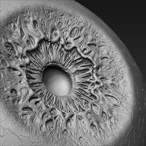
Iris is a pigmented, round, contractile membrane of the eye, suspended between the cornea and lens and perforated by the pupil (Fig. 1). It regulates the amount of light entering the eye (Online Dictionary). According to (David J. Pesek, 2010), the eyes are connected and continuous with the brain’s Dura mater through the fibrous sheath of the optic nerves, and they are connected directly with the sympathetic nervous system and spinal cord. The optic tract extends to the thalamus area of the brain. This creates a close association with the hypothalamus, pituitary and pineal glands. These endocrine glands, within the brain, are major control and processing centers for the entire body. Because of this anatomy and physiology, the eyes are in direct contact with the biochemical, hormonal, structural and metabolic processes of the body. This information is recorded in the various structures of the eye, i.e. iris, retina, sclera, cornea, pupil and conjunctiva. Thus, it can be said that the eyes are a reflex or window into the bioenergetics of the physical body and a person’s feelings and thoughts (David J. Pesek., 2010). There are a lot of arguments between iridologists (iridology’s practitioner) and the medical’s practitioner. Due to this argument, numerous studies done by the medical’s practitioner found that the diagnosis done by the iridologist upon the patient is not accurate (Allie Simon et al, 1979). However the study on relationship diseases to iris changes, still continuing for example the studied done on Ocular complication of adult rheumatoid arthritis don by S.CReddy and U.R.K.Rao in 1996 found that the mean duration of the arthritis and the mean duration of seropositivity were found to be significantly higher in patients with ocular (pigmented organ in eye) complication (S.CReddy et al, 1996). Another study done on bilateral retinal detachment in acute myeloid Leukemia by (K Pavithran et al., 2003), found that ocular manifestations are common in patient with acute Leukemia. This can result from direction infiltration by neoplastic cells of ocular tissues, including optic nerve, choroid, retina, iris and ciliary body, or secondary to hematology abnormalities such as anemia, thrombocytopenia, or hyperviscosity states or retinal destruction by opportunistic infection (K Pavithran et al., 2003). The history of Iridology study on iris was done by the physician Philippus Meyens in 1670 in a book explaining that the features of the irid called Chromatica Medica. In that book he wrote that the eye (iris) contains valuable information about the body. In 1881 a Hungrarian physician, Dr. Ignatz Peczley who is claimed as the founder of modern Iridology wrote a book “Discoveries in the Field of Natural Science and Medicine, a guide to the study anddiagnosis from the eye.” He introduced the first chart of the iris explaining zone in the iris. The idea of his study on iris, begun when he was a child, he was accidentally found the Owl with broken leg. He found a dark scar in the Owl’s iris that scar turned white as the leg healed (Sandy Carter, 1999). The objective of this chapter is to explain how the presence of cholesterol in blood vessel can be detected by using iris recognition algorithm. This method used the John Daugman’s and Libor masek’s iris recognition methods and extends the study of eyes pattern to other application and in this case, the alternative medicine that is iridology. Based on the iris recognition methods and iridology chart, a MATLAB program has been created to detect the present of cholesterol in our body. However, further analysis must be done in order to know the exact range or level of cholesterol in blood vessel.
Ridza Azri Ramlee, Khairul Azha and Ranjit Singh Sarban Singh Universiti Teknikal Malaysia Melaka (UTeM),
Malaysia
Download full abstract: 21767

Leave A Comment