Abstract
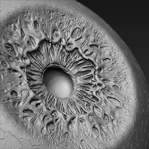
The study takes advantage of several new breakthroughs in computer vision technology to develop a new mid-irisbiomedical platform that processes iris image for early detection of heart-disease. Guaranteeing early detection of heart disease provides a possibility of having non-surgical treatment as suggested by biomedical researchers and associated institutions. However, our observation discovered that, a clinical practicable solution which could be both sensible and specific for early detection is still lacking. Due to this, the rate of majority vulnerable to death is highly increasing. The delayed diagnostic procedures, inefficiency, and complications of available methods are the other reasons for this catastrophe. Therefore, this research proposes the novel IFB (Iris Features Based) method for diagnosis of premature, and early stage heart disease. The method incorporates computer vision and iridology to obtain a robust, non-contact, nonradioactive, and cost-effective diagnostic tool. The method analyzes abnormal inherent weakness in tissues, change in color and patterns, of a specific region of iris that responds to impulses of heart organ as per Bernard Jensen-iris Chart. The changes in iris infer the presence of degenerative abnormalities in heart organ. These changes are precisely detected and analyzed by IFB method that includes, tensor-based-gradient(TBG), multi orientations gabor filters(GF), textural oriented features(TOF), and speed-up robust features(SURF). Kernel and Multi class oriented support vector machines classifiers are used for classifying normal and pathological iris features. Experimental results demonstrated that the proposed method, not only has better diagnostic performance, but also provides an insight for early detection of other diseases.
21 July 2017

Leave A Comment