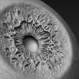
Besides Physical test normally, early detection on the condition of the body by using the image processing of iris is an alternative method to observe the health of human’s body, especially the internal organ of the body. This paper uses Dr. Bernard Jensen’s chart of iris as reference, in which part and how deep is the damage happens in the tissue of iris. Organ disorder is represented by the form of broken tissue of iris. The broken tissue usually seems to be like a hole in certain area in the iris. In this paper, the instrumentation for data mining uses video camera and the software that will be developed uses Visual Basic on image processing programming. In the image of eye, the region of interest is only on the iris, and it will be grabbed by using circle and line equations.The area of Liver organ lies on 07.15 – 07.45 in the third Quadran. After wards, this slice of image is prepared for image processing system. The method that is going to be used in this paper is grey level, enhancing and sobel operator. Then, the output of the system will be compared with physical test to measure the precision on detecting the problem on Liver organ.
CONCLUSION
From the research that has been taken several conclusions, namely:
1. The system as a whole, starting from the instrumentation that was tried to be developed through a video camera, image processing method with SOBEL multiplier matrix, is able to work well, which is 84% correct to detect any interference in the liver organ that is represented in the form of “broken tissue” in in iris imagery.
2. The image processing method can be used to determine the location of the iris image and the position of the liver organ. The image processing method is able to detect whether or not there are holes in the iris ROI image.
SUGGESTIONS
1. New image processing methods need to be developed for to accurately detect (100%) the position of the liver organ.
2. It is also necessary to develop a realtime data retrieval system so that data can be directly tested directly.
3. The detection process can also be developed in other organs and supported by medical data as well.
4. From this study the author was inspired to explore Iridology as a Pre-Diagnostic medium for doctors so that in the future it may be possible to do Data Mining in greater terms about the reality of the relationship between the presence of “broken tissue” signs in the iris.
